Research Zone

Deptt. Tuberculosis & Chest Diseases
Govt. Medical College, Patiala
On Going Research Projects
1. To study the outcomes of Pleural Effusion in patients attending TB & Chest Hospital Patiala
2. To study clinic radiological profile of Interstitial Lung Diseases
3. To study clinic radiological profile of Allergic Broncho Pulmonary Aspergillosis
4. To study the knowledge of prescriptions of Anti Tubercular Treatment among private practioners
5. Clinico radiological spectrum of Pulmonary & Extra Pulmonary TB in HIV Patients
6. To study drug sensitivity pattern in patients of MDR TB
7. Questionaire based study on level of satisfaction in patients admitted in Chest & TB Hospital, Patiala
Clinical and Radiological Spectrum of Pulmonary Lesion in HIV Positive Patients
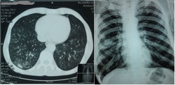
Even in HIV infected patients, pulmonary tuberculosis is the most common oppurtunistic infection. Typical chest X-ray findings like upper lobe infiltrates with cavitation are less common as compared to atypical findings like bilateral infiltrates (especially lower zones)
with or without cavitation, a miliary pattern or involvement of different groups of lymphnode occurs.
This study is being conducted on HIV positive patients with tuberculosis (both pulmonary ‘PTB’ and extrapulmonary tuberculosis ‘EPTB’) presenting to Department of Chest and TB, Govt. medical college, Patiala. 26 patients were studied out of which 13(50%) were of PTB and 13(50%) were of EPTB. EPTB cases consisted of, 7(54%) cases of hilar lymphadenopathy, 4(31%) of pleural effusion, one case (8%) each of tubercular meningitis and abdominal kochs. CD4 cell count was less than 300 in 17(65%) and more than 300 in 4(15%). CD4 count was not known in 5(20%) patients. Chest xray revealed lower zone involvement in 6(23%), bilateral extensive disease in 9(35%), hilar lymphadenopathy in 7(27%) and pleural effusion in 4(15%) of patients and none showed typical upper zone involvement. Sputum for AFB was negative in 18(69%), positive in 8(31%) of patients.
Conclusion- The results from above study clearly shows that atypical pattern on chest xray in tuberculosis patients with HIV in form of lower zone involvement, different groups of lymphnode enlargement is more common. Secondly greater number of patients with HIV having lesion on chest xray are acid fast bacillus (AFB) smear negative.
Utility of Computed Tomography Guided Percutaneous Transthoracic Fine Needle Aspiration Cytology in Different Lung Lesions
Percutaneous transthoracic fine needle aspiration cytology is a well established diagnostic method used in the cytological evaluation of both neoplastic and inflammatory conditions of lung . CT offers detailed anatomical display of all thoracic structures and CT guided FNAC of suspicious lung masses is widely accepted simple diagnostic method of relatively low cost used to establish diagnosis of lung and chest lesion.
A hospital based descriptive study is being done to know the pathological spectrum of thoracic lesions obtained from computed tomography guided percutaneous transthoracic fine needle aspiration.
The clinical, radiological and cytological data of 70 patients were studied who underwent CT guided FNAC from July, 2010 to July, 2011 and the study is still going on.
Cytological examination showed that 22 cases were malignant and 48 cases were non malignant. Provisional diagnosis based on radiological and clinical findings were 38 and 32 cases of malignant and non malignant lesions respectively.
Post procedure complications were pneumothorax in two cases which was self resolving did not require any intervention and hemoptysis in one case which also did not require any active management.
CT guided fine needle aspiration cytology (FNAC) is a simple and safe procedure with high diagnostic accuracy for the diagnosis and cell typing of lung lesions especially where this facility exists. Though complications are rare, pneumothorax, perilesional hemorrhage, hemoptysis and chest pain are occasionally encountered, but rarely require active management.
Aspergillus Hypersensitivity in Patients with Chronic Obstructive Pulmonary Disease
Allergic bronchopulmonary aspergillosis (ABPA) caused by hypersensitivity to fungus aspergillus (most commonly Aspergillus fumigates) primarily complicates the course of asthma. There is a possible risk that patients with chronic obstructive pulmonary disease (COPD) can also develop Aspergillus hypersensitivity (AH)
Aim and Method
The aim of this study conducted in the Deptt. of Chest And TB, Govt. Medical College, Patiala was to evaluate the prevalence of AH in patients with COPD. One hundred subjects with confirmed diagnosis of COPD and one hundred normal healthy volunteers were subjected to aspergillus skin test. Patients were said to have AH if they demonstrated immediate cutaneous hyperreactivity to Aspergillus fumigates.
Outcomes Of Pleural Effusions: Dept of Pulmoanry Medicine
The study was conducted on 51 patients who presented to Dept. of Chest And TB, Govt. Medical College, Patiala with chest radiograph suggestive of pleural effusion. Diagnostic/Therapeutic aspiration done and fluid was sent for complete analysis. Patients were treated after reviewing the reports and treatment outcome was studied.
Out of 51 patients 27(53%) had right sided pleural effusion,19 had left sided pleural effusion and 5(10%) had bilateral pleural effusion. Out of these 3(9%) were Human Immunodeficiency Virus reactive also.
Fluid was neutrophilic in 14(27%) cases and lymphocytic in 27(53%) and 6(12%) were malignant in nature. Ziehl Neelson stain was positive for Acid fast bacilli in 7(14%) patients. None showed growth on culture. One showed cyst of Entamoeba Hystolitica. 15(29%) were subjected to Intercostal tube drainage, out of which 9(60%) were extubated after lung expansion within a week while 3 patients were referred for surgical intervention. One patient died and two were lost in follow up. 31 patients were started on Antitubercular treatment.
Thus, it is concluded that diagnostic and therapeutic aspiration in pleural effusion is easy, cheap and can be done on outdoor patients. It helps in reaching at proper diagnosis quickly and effective medical treatment. Ziehl Neelson stain of fluid has poor yield in diagnosis of tubercular pleural effusion but if the treatment is started on the basis of clinical history, cytology, serology and biochemical analysis, results are quite good. The need for surgical intervention is alsmost negligible which decreases the morbidity and mortality to a large extent.
